Imaging Center
With Doctors W. Gibby, A. Gibby, C. Gibby, and J. Gibby
Blue Rock Imaging utilizes high-tech equipment and advanced techniques to create clear and accurate images of bones, soft tissues, organs, blood vessels, arteries, and the nervous system.
Imaging Services

MRI
A test that produces

CT Scan
A scan used to reveal damaged organs, ear problems, fractures, and more.
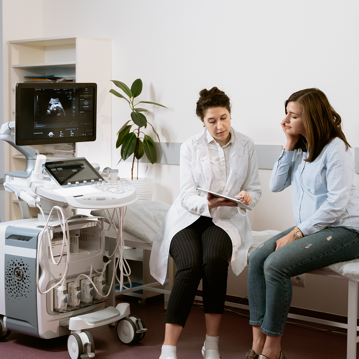
Ultrasound
High-resolution ultrasounds use sound waves to monitor inside your body.

X-Ray
X-ray radiation passes through part of the body to produce an image.

3D Mammography
Breast Cancer can be screened using mammograms, ultrasounds, MRIs and biopsies.

Fluoroscopy
Fluoroscopy is used in various examinations to diagnose or treat patients.

Angiography
The use of injected dye to increase the contrast of arteries in an imaging test.

Dexa
Bone density and body composition scans.
MRI
Duration: 15 - 90 minutes
What is an MRI?
An MRI is a test that produces images of internal organs and structures virtually anywhere in the body utilizing a strong magnetic field and interaction with radio waves.
An MRI can be used to detect and diagnose tumors and diseases. It may also be used to provide other information that cannot be seen using other medical imaging methods.
What should I expect from an MRI?
While every person's experience will be slightly different, here is a basic idea of what to plan for:
- Arrive early
- You will be asked to remove any clothing containing metal and all jewelry. You will be provided metal free clothing to change into such as a gown with shorts or pants. Lockers are available to safely store all personal items and electronics.
- Our certified tech will provide you with some brief instructions and help prepare you.
- The test can be noisy. Ear plugs will be available if desired.
- Lie still during the duration of the test.
- You're done!
Types of MRI
- Head/Neck Brain, Sella, IACs, Orbits, MRA Brain, MRA, Carotids, Soft Tissue Neck, Thyroid, and Parathyroid.
- Spine Cervical, Thoracic, Lumbar, and Sciatic N. Survey.
- Chest Brachial Plexus, Chest, Heart, Breast, and Contrast MRA.
- Abdomen/Pelvis Abdomen, Cholangiogram, Liver, Adrenal, Kidney, Pancreas, Pelvis, Hip, MRA, and MRV.
- Upper Extremity Shoulder, Elbow, and Wrist.
- Lower Extremity Knee, Ankle, and Foot.
- FMRI
X-Ray
What is an X-ray?
A digital X-ray is a test that produces images by passing X-ray radiation through a part of the body.
Types of X-rays include: Abdomen, ankle, cervical spine, chest, clavicle, coccyx, elbow, facial bones, femur, finger, foot, forearm, hand, humerus, knee, lumbar spine, nasal bones, orbits, pelvis, ribs, sacrum, shoulder, sinus, skull, sternum, thoracic spine, tib/fib, toe, and wrist.
Dexa
Bone Density Scan
Check in: 10-15 minutes
Scan Time: 15 minutes
Avoid Calcium supplements 24 hours before scan.
Did you know? Nearly 1 in 5 women and 1 in 20 over the age of 50 are affected by osteoporosis. A bone density scan can tell your primary care doctor invaluable information!
Fluoroscopy
What is fluoroscopy?
Fluoroscopy is a study of moving body structures and is often compared to an X-ray "movie." A continuous X-ray beam is passed through the body part being examined. The beam is transmitted to a TV-like monitor so that the body part and its motion can be seen in detail.
Fluoroscopy is used in a wide variety of examinations and procedures to diagnose or treat patients. Some examples are:
- Abscessogram
- Arthrogram
- Barium Swallow
- BE Double Contrast
- BE Single Contrast
- Catheter Check
- Hysterosalpingogram
- Lumbar Puncture
- Nathan Green Small Bowel
- Small Bowel Follow-through
- Upper GI
- VCUG
CT Scan
Duration: Usually about 30 minutes
What is a CT Scan?
A CT Scan reveals torn or damaged organs, inner ear problems, broken bones, vertebral bone damage, etc. that cannot be diagnosed using regular x-rays.
Is there anything I need to do to prepare for a CT Scan?
Follow your doctor's instructions. Before your scan you will change so there is no metal on your body (a gown is provided if your clothes have any metal). You will lay on a long cushioned panel that moves through a circular opening. Tubes will move around the panel and x-ray your body.
Types of CT Scans
- Full-body screening
- Virtual colonoscopy
- Head and neck
- Spine
- Chest and abdomen
- CT Angio
X-Ray
What is an X-ray?
A digital X-ray is a test that produces images by passing x-ray radiation through a part of the body.
Types of X-rays include: Abdomen, ankle, cervical spine, chest, clavicle, coccyx, elbow, facial bones, femur, finger, foot, forearm, hand, humerus, knee, lumbar spine, nasal bones, orbits, pelvis, ribs, sacrum, shoulder, sinus, skull, sternum, thoracic spine, tib/fib, toe, and wrist.
3D Mammography
What is a 3D mammogram?
A non-invasive X-ray creates 3D images of your breast that are used to screen for breast cancer and other abnormalities. The images will be reviewed by our board-certified radiologists and a report will be sent to you or your doctor.
When should you get a mammogram?
When you reach the age of 40, a yearly mammogram is recommended. You should also consider getting checked if you have a family history of Breast Cancer or notice changes in your breasts.
What should you expect?
Knowing what’s going on with your breasts is easier than you think with a 3D mammogram. It’s no surprise a 3D scan is better at detecting breast abnormalities than 2D pictures. Our Hologic machine offers enhanced accuracy for women of all ages and all levels of breast density. The process is very simple:
- In a private room, you'll undress from the waist up and put on a gown. It's best if you don't wear any deodorant, powder, or glitter lotion.
- One of our radiologic technologists will meet you in a private mammo suite.
- While standing, the technologist will position you in front of the machine and place one of your breasts on the machine.
- The machine will start and a robotic arm with arc over your breast and take multiple x-ray images. The compression may cause a bit of pressure but it shouldn't be painful.
- After one breast is imaged, the same process is repeated with the other breast.
- You're all done!
Breast Cancer Screening Methods
- 3D Mammogram
An x-ray of the breast is performed for either screening or diagnosis. - Breast Ultrasound
After a mammogram, a machine uses sound waves to look inside the breast to determine whether the lump previously discovered is a cyst or a tumor if needed. - Breast MRI
An MRI provides detailed pictures of areas in the breast using a magnet linked to a computer. - Breast Biopsy
Using our high-resolution ultrasound, tissue or fluid is removed from the breast and sent to pathology.
Pelvic Congestion Syndrome
How is pelvic congestion syndrome treated?
Gonadal vein embolization is a minimally invasive treatment for pelvic congestion syndrome. The goal is to close off faulty veins so they can no longer enlarge with blood, thus relieving pain and symptoms of pelvic congestion syndrome.
CT Scan
What is a CT Scan?
A CT Scan reveals torn or damaged organs, inner ear problems, broken bones, vertebral bone damage, etc. that cannot be diagnosed using regular x-rays.
Is there anything I need to do to prepare for a CT Scan?
Follow your doctor's instructions. Before your scan you will change so there is no metal on your body (a gown is provided if your clothes have any metal). You will lay on a long cushioned panel that moves through a circular opening. Tubes will move around the panel and x-ray your body.
Types of CT Scans
- Full-body screening
- Virtual colonoscopy
- Head and neck
- Spine
- Chest and abdomen
- CT angio
High-Resolution Ultrasound
How does an ultrasound work?
An object called a transducer is pushed onto the patient's skin. The transducer sends small pulses of high-frequency sound waves into the body. When the sound waves hit an object, they bounce back or echo. The transducer measures these bounces and uses them to project an image onto the monitor.
Ultrasound may be used for:
- Gallbladder disease diagnosis
- Evaluating blood flow
- Imaging of the uterus and ovaries
- Monitoring a developing baby's health
Is there anything I need to do to prepare for an ultrasound?
Patients will need to remove any jewelry from the area being examined and may need to change into a gown.
Angiography
What is angiography?
A test using X-rays, MRI scans or CT scans to detect blockages in the arteries. Dye (contrast) is injected into a blood vessel before the images are taken. The contrast helps the arteries show up more clearly on the test images.
Arteriogram:
- Cerebral/Carotids
- Renal
- Run-off/Lower Extremity
- Visceral
- Av Fistula/Graft
- Aortogram
- Pulmonary
- Subclavian Angiogram
Venous Access:
- Picc Line
- Central Line
- Portacath
Visit Blue Rock Imaging Today

Visit Blue Rock Imaging Today
Address
3152 N University Ave, Provo, UT 84604
Suite #100
Phone Number
Hours
Monday - Thursday
7:30am - 6pm
Friday
7:30am - 5:00pm